Wednesday, August 15, 2018

AN 80+ YEAR OLD SECRET REVEALED !

In 1936 April, some 82 years back, Journal of Biochemistry published an article on metabolism of glutamic acid in brain. Hans Weil-Malherbe, the author of this long forgotten research, had looked into metabolism of both L- and D-glutamic acid in brains of two animal species (Guinea pig and rat) in a review.

Major points of this Newcastle scientist's review are summarized here as follows:

1. Glutamic acid is different from all other amino acids, as it is the only amino acid oxidized in brain.

2. L-glutamic acid acts as a substrate of brain respiration.

3. Although not very efficient, L-glutamic acid can maintain brain respiration.

4. D-glutamic acid, a non natural isomer cannot be oxidized in brain.

5. If D-glutamic acid appear with or without glucose in brain, slight reduction of brain respiration occurs.

scanned image of page 668
6. Although, brain slices only attacks natural isomer (L-Glutamic acid), brain extracts not only attacks both D- and L- isomers, but attack D-Glutamic acid preferentially. (Brain extracts contain 'Glutamic acid deaminase enzyme). Under both aerobic and anaerobic conditions, brain extracts oxidize L-isomer slowly when compared with D-isomer.

7. Tumor slices as well as ox kidney slices attack both L- and D-isomers at almost same rate.

8. Glutamic acid is the only amino acid oxidized by tumor.

9. Differences of reactions between two tissues (brain and kidney) were thought to be due to differences in chemical composition; mainly accounted by high lipoid content in brain, that was thought to hold enzyme (Glutamic acid deaminase) more tightly.

10. Properties of glutamic acid deaminase in brain:

(a) O2 uptake
(b) Ammonia production
(c) enzyme is specific to D-Glutamic acid

11.

Table XII. Oxygen uptake and ammonia formation with extract of dry brain powder after ether and alcohol extraction. Powder1 of Table XI repeatedly treated with abs. alcohol at room temp. Alcohol removed in vacuo over P205. Extracted with 15 parts of M/100 veronal buffer (PH 8-2) and centrifuged. Each vessel contained 2 ml. of the extract. Duration of the experiment: 6 hours.

Substrate added
O2 uptake (micL)
Extra O2 (micL)
NH3 formation (micL)
Extra NH3 (micL)
Ratio
O2:NH3
0
33.5
-
48.9
-
-
M/50 L-alanine
34
+0.5
46.6
-2.3
-
M/50 DL alanine
32
-1.5
48.3
-0.6
-
M/50 DL valine
34
+0.5
50.6
+1.7
-
M/50 L-glutamic acid
37
+3.5
45.4
-3.5
-
M/50 D-glutamic acid
58
+24.5
73.6
+24.7
1 : 1.01

12. L-glutamic acid (only amino acid oxidized in brain), is oxidized first into alpha-keto glutaric acid and NH3, then into H2O and CO2.  The enzyme responsible for the oxidation of 1( + )-glutamic acid to oc-ketoglutaric acid and NH3 does not attack d(-)-glutamic acid so long as it is bound in the cell or to some constituent of the cell, probably a lipoid. In solution however the specificity is changed and d(-)-glutamic acid alone is oxidised.


WHAT DIDN'T HE POINT OUT FROM HIS FINDINGS?

  • HIGHLIGHTED FIGURES CLEARLY SHOW PRESENCE OF D-GLUTAMIC ACID IN BRAIN SUBSTANTIALLY INCREASE OXIDATIVE STRESS AS WELL AS AMMONIA FORMATION.
  • THIS INCREASES A THEORETICAL POSSIBILITY OF DEGENERATIVE DAMAGE OF BRAIN TISSUES IF A LONG TERM EXPOSURE TO D-GLUTAMIC ACID OCCUR.
  • MAJORITY OF COMMERCIALLY USED MSG  AT PRESENT CONTAINS D-GLUTAMIC ACID. AND IT IS PRODUCED IN BILLIONS OF TONS ANNUALLY TO BE ADDED TO OUR FOODS. AT THE SAME TIME, DEGENERATIVE BRAIN DISORDERS ARE SKYROCKETING. SEE A LINK?

NOTE; This article was published in 1936. Commercial use of MSG as a flavor enhancer was first started in early 1940's. Further, until 1957, commercially produced MSG mainly contained L-glutamic acid. It was only after that year, D-glutamic acid dominated the market. So, it's no wonder that the author of this study had not seen any relevance of drawing these conclusions. But it carries a great importance today - as we are eating this dangerous D-glutamic acid in billions of tons per year.

Source:

Studies on brain metabolism
The metabolism of glutamic acid in brain
Hans Weil-Malherbe . 1936 Apr; 30(4): 665–676.


Tuesday, August 14, 2018

D-GLUTAMATE CANNOT PARTICIPATE IN MITOCHONDRIAL PUMPS

Mitochondria are the power houses of cells. Without energy produced by them, cells cannot efficiently function. Glutamate H+ co transporters at mitochondrial membrane helps to maintain optimal pH in these power houses. "However, these pumps can only utilize L-type glutamate", a Cambridge University molecular research team explains.

Processed Mono Sodium Glutamate (MSG), dominating flavor enhancer in modern food industry contains D-glutamate. Though, exact mechanisms of D-glutamate processing inside body are less clear, over consumers of D-type dietary glutamate sources have to be cautious, according to current single cell study.

Image result for mitochondria diagram

Source:

Identification of the mitochondrial glutamate transporter:
bacterial expression, reconstitution, functional characterization,
tissue distribution of two human isoforms*
Giuseppe Fiermon‡,t eLuigi Palmier‡i, Simona Todis‡c,o Gennaro Agrimi‡,
Ferdinando Pmalieri‡¶ and John E. Walk§e¶r
JBC Papers in Press. Published on March 15, 2002 as Manuscript M201572200

D-SERINE : AN EXCEPTIONAL D-AMINO ACID IN BRAIN

"In contrast to L-isomer domination of amino acids among mammalian tissues, D-serine functions as a neurotransmitter in brain", a study reveals. It acts on high affinity site on NMDA (M-methylD-aspartate) receptor. It's involved in an important role in glial cell neurotransmission or gliotransmission. Serine racemase, the enzyme which produces D-serine is also found widely distributed in brain tissues.

Both NMDA receptors and astrocytes are key players of glutamate linked metabolism in neural tissues. However, exact role of D-serine in brain glutamate metabolism is yet to be determined.


d‐Serine localizes to neurons and astrocytes in the brain. Staining for d‐serine was performed in pyramidal neurons of layer V of the cerebral cortex and in astrocytes in the corpus callosum of a P9 rat. The lower panels depict double‐labeling immunofluorescence for d‐serine (labeled for SR in the original publication) and a neuronal nucleus marker (NeuN) in layer VI of the cerebral cortex of a P9 rat. Reproduced with slight modifications from Kartvelishvily et al.

Source: 

dAmino acids in the brain: dserine in neurotransmission and neurodegeneration.             Herman Wolosker, Elena Dumin, Livia Balan,Veronika N. Foltyn.
FEBS Journal.
Dec 31, 2010. Pages 3514-3526

D-AMINO ACIDS IN PROCESSED FOODS : WHAT HAPPENS INSIDE THE BODY?

Food processing makes two major changes in plant proteins -

  • Racemization of all L-amino acids to D isomers
  • Formation of cross linked amino acids (ex. lysinoalanine)
Racemization is depend on pH, time and temperature exposed. Conversion rates of isomers differ. However, D-amino acids present in food comes from both processed as well as from natural foods. Latter sources include plants, micro organisms and marine invertebrates. Whatever the source, racemization impairs nutrition quality and digestibility.

Some D-amino acids can be used in animal bodies.
  • D-methionine: nutritional source of L-isomer
  • D-phenylalanine: nutritional source of L-isomer
Image result for d amino acid

Others either useless or cause harmful effects.
  • high concentrations of D-tyrosine fed to mice, inhibited their growth rates
  • D-serine: cause histological changes in rat kidneys
  • Lysinoalanine: cause histological changes in rat kidneys
  • D-lysine: no nutrional value
  • D-cysteine: no nutrional value
Humans expose to these D-amino acids, specially from diets high in processed foods. but their safety, physiological roles and potential harmful effects are grossly under studied.

Source:

Chemistry, Nutrition, and Microbiology of d-Amino Acids. Mendel Friedman. Western Regional Research Center, Agricultural Research Service, U.S. Department of Agriculture, 800 Buchanan Street, Albany, California 94710. J. Agric. Food Chem.199947 (9), pp 3457–3479

YOUNGSTERS CANNOT HANDLE D-AMINO ACIDS

"Younger animals have lower amounts of D amino acid oxidases and consequently higher concentrations of free D-amino acids compared to adult animals. If the ingested D-amino acids are not metabolized by these enzymes, they will accumulate in the tissues and may provoke serious damage leading to suppression of the synthesis of other essential enzymes and inhibition of the growth rate of the animals", an Italian research team says.

Amino acids can be exist in L or D forms of photo isomers - that means non superimposable  mirror images of same molecule. Usually, L amino acids naturally exist in animal bodies in abundance. D isomers often comes from synthetic sources. When an animal ingests a D amino acid, it absorbs from intestine and is transported through blood stream to organs such as liver and kidneys. These organs either have or produce D amino acid oxidases, to detoxify D amino acids.

Image result for d amino acid

Under mentioned process of D amino acid oxidase or Aspartate oxidase produces ammonia and hydrogen peroxide as toxic byproducts. They increase oxidative stress, hence the need for antioxidants are raised.

If mother ingests these free D-amino acids in her pregnancy, young rats showed increased production of D-amino acid oxidases. "Specifically, when a mother rat ingests D-Ala or D-Asp during pregnancy and suckling, an increase in D-amino acid oxidase or D-aspartate oxidase is observed in the liver and kidneys of the baby rats", Dr. Aniello, the lead scientist of current study explains.

Source:

Biological role of D-amino acid oxidase and D-aspartate oxidase. Effects of D-amino acids.

December 25, 1993The Journal of Biological Chemistry268, 26941-26949.


CAMEL MILK PROTECTS TESTES AGAINST MSG DAMAGE

A research team made of Egyptian and Saudi Arabian scientists had examined the effects of camel milk and vitamin E on it's ability to protect testicular tissues and male endocrine derangement from toxic MSG effects.

MSG caused following toxic effects on testes:

  • MSG induced testicular apoptosis (death of testicular cells).
  • At the molecular levels, MSG reduced gene expression of testicular hormones and reduced receptors of androgen, Luteinizing Hormone (LH) and follicular stimulating hormone (FSH).


They had found superior ability of camel milk (CM) to vitamin E in exerting following effects:

  • CM normalized the decrease in serum levels of testosterone, Luteinizing Hormone (LH), sperm profiles and testicular antioxidant activities that were decreased by MSG.
  • All altered genes were normalized and upregulated in presence of CM when compared with vitamin E.
Current study team suggests usage of camel milk as a therapy against MSG used in food industry.

Image result for camel

Sources:


El-Sawy HBI, Soliman M, El-Shazly S, Abdel-Maksoud Ali H. Protective effects of camel milk and vitamin E against monosodium glutamate induced biochemical and testicular dysfunctions. PN [Internet]. 29Mar.2018 [cited 14Aug.2018];20(1):76-5.


MSG – “AD LIBITUM” DIELEMMA


It had been repeatedly observed that very high doses of MSG in “ad libitum” method is needed to produce neuro degenerative damage in brains of experimental animals.

(“FREEDICTIONARY” defines “ad libitum” feeding as ‘food available at all times with the quantity and frequency of consumption being the free choice of the animal’.)

This is complete opposite to mice models who received MSG by forced oral route and by subcutaneous route where brain damage could be induced by 1/10 to 1/20 doses (2 g/kg to 4 g/kg) of MSG that was required to induce same lesions with “ad libitum feeding” (20.9 g/kg to 45.5 g/kg).

Related image

“The cause for this difference lies in the route of administration”, experts say. When MSG is given in oral route, body calls in multiple defenses to keep blood glutamate levels stable in order to prevent MSG toxicity. These defenses are;

1.      Metabolizing glutamate in gut.
2.      Extrusion from brain by active blood-brain transport systems.
3.      By local brain mechanisms regulating glutamate uptake and metabolism in brain.

These systems can buffer glutamate to maintain extracellular glutamate level (glutamate level outside brain cells) stable.

“However, mechanism of MSG neurotoxicity often attributed to a prolonged increase in extracellular glutamate concentrations in brain”, a recent review on toxic effects of MSG explain.


Sources:

1.      Olney JW. Excitotoxic amino acids: Research applications and safety implications, In Flier, L J, Garratini, S, Kare, M R Reynolds, W.A and Wurtman, R.J (Eds.), Glutamtc Acid Advances in Biochemistry and Physiology, Raven. New York, 1979.

2.      A Studies On Monosodium L- Glutamate Toxicity In Animal Models- A Review. Kumar Ganesan1, Kumeshini Sukalingam1, Kanaga Balamurali1, Siti Radziah Bt. Sheikh, Alaudeen1, Kumar Ponnusamy1, Indang Ariati Ariffin, Sharmila Banu Gani. International Journal Of Pharmaceutical, Chemical And Biological Sciences, 2013, 3(4), 1257-1268.


EVEN LOW DOSES OF MSG DAMAGE LIVER CELLS

"Liver that account for detoxifying toxic material present in food, hence faces first challenges of food associated toxic hazard, is liable to Mono Sodium Glutamate (MSG) induced liver damage even at very low doses", Dr. Eveka who looked into matter with an animal experiment says.

The team had used two strengths of very low doses of MSG mixed in Growers' mash daily for 42 days. Histological changes in livers were examined after 42 days. Observed changes include:

  • Dilatation of central vein with lysed red cells
  • Cyto-architectural distortions of hepatocytes
  • Atrophic and degenerative liver changes 
Image result for central vein of liver cells

All examined biochemical parameters were elevated:
  • Aspartate aminotransferase (AST)
  • Alanine aminotransferase (ALT)
  • Total Protein
  • Albumin
Further, animals who exposed to higher MSG dose (.08 mg/kg) showed all changes more than mice who exposed to low MSG dosage (.04 mg/kg).

This study highlights two important facts;

1. It had used very low dosages (compare with usual doses used in MSG animal experiments - subcutaneous or oral 2 mg/kg or 4 mg/kg) in mixed feeds (NOT in forced feeds)

2. It's clear that even at these very low doses liver cells could be damaged.

As liver cells are subjected to first pass circulation, they are very vulnerable to toxins absorbed through intestine from food. That means liver faces first to food toxins and are liable to hepatocyte destruction. It is only when liver cells are significantly damaged,  blood levels of MSG rise and rest of the body expose to toxic MSG effects. 

Also read on higher doses of MSG needed in 'ad libitum' exposure to cause neuro toxic effects.
https://hapicureclinic.blogspot.com/2018/08/msg-ad-libitum-dielemma.html

Source:


Annals of Medical and Health Sciences Research – January 2011 – Vol. 1 N0.1  Histochemical Studies of the Effects of Monosodium Glutamate on the Liver of Adult Wistar Rats Eweka AO, Igbigbi PS, and Ucheya RE.


Monday, August 13, 2018

GARLIC WORKS AGAINST MSG

"Allicin, active ingrediant present in chopped garlic properties can attenuate hepato-toxic effects of Mono Sodium Glutamate (MSG) in experimental mice models", Dr. Elbezzoumy of Alexandria University, says.

During their experiment, they had exposed two groups of mice to subcutaneous 4 mg/g MSG doses for 45 days. Only one group was additionally provided 10 g/kg diet of garlic extract (1%).

Garlic extract was capable of reducing;

  • liver oxidative stress
  • liver tissue damage
  • hyperglycemia
  • hyperlipidemia
The researchers attribute these benefits mainly to supportive role of allicin in regeneration of glutathione system (a potent anti oxidant defense mechanism of human body) components. 

Related image

Source:

1. 
J B B 1(1) 2010 © INTERNATIONAL SCIENCE PRESS Toxic Effects of Monosodium Glutamate on the Hepatic Tissue Damage of Rats and the Beneficial Role of Garlic Extract: Involvement of Oxidative Stress, Hyperlipidemia and Hyperglycemia ASHARF M. ELBESSOUMY1 , DOAA A. GHAREEB1 AND ASHRAF A. KHALIL

MSG AND HIDDEN HUNGER

Reduction of oxidative stress of nerve cells is a very important step in lowering MSG induced nerve damage. There are very efficient anti oxidant systems exist in human body. They include:

1. Vitamin C
2. Vitamin E
3. Glutathione - Needs B complex vitamins for regeneration.

Image result for vit e rich food

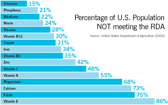
U.S. Department of Agriculture, lists U.S. citizens'hidden hunger' or micro nutrient deficiencies as below.

Image result for usa micronutrient deficiency list

According to which, 86% of people are vitamin E deficient, while vitamin C deficiency is prevalent in 48% of population. This again shows vulnerability of nearly entire U.S. population to MSG induced neuronal damage.

Sources:

1. Traynelis, S. F., Wollmuth, L. P., McBain, C. J., Menniti, F. S., Vance, K. M., Ogden, K. K., … Dingledine, R. (2010). Glutamate Receptor Ion Channels: Structure, Regulation, and Function. Pharmacological Reviews62(3), 405–496. http://doi.org/10.1124/pr.109.002451

2. https://www.nal.usda.gov/fnic/vitamins-and-minerals


WHY AT LEAST 68% OF U.S. POPULATION ARE VULNERABLE TO MSG TOXICITY?

Glutamate act as the most abundant neurotransmitter in brain. but, as it is an excitatory neurotransmitter, it's extracellular concentration (glutamate concentration outside the brain cells) are very tightly controlled. The main regulatory mechanism involves astrocytes (the star shaped cell in the diagram), which can convert GLU (Glutamate) to GLN (glutamine) and then release GLN to extracellular space. Then glutamatergic neurons capture these GLN molecules and convert them back to GLU, which is now released into synaptic space where GLU exerts it's function as a neurotransmitter. As soon as neural message is transmitted, GLU molecules are efficiently recycled by astrocytes.


Image result for Astrocyte recycling of glutamate


What happen if this mechanism fails (for an example, in a chronic high dose exposure to MSG) ? Then excitatory (means it can fire neurons - neuron firing is exactly needed to neural message transmission, but if this happens out of control, the neurons would die) GLU pile up at synaptic space. They act on GLU receptors.

There are two major types of GLU receptors: ionotropic and G protein coupled receptors. Ionotropic receptors are of three sub types; NMDA, AMPA and Kainate. Out of these, NMDA and AMPA receptors are often co located, while Kainate receptor distribution in brain is limited to few specific areas. Basically, these ionotropic type of receptors dominate in cortex, basal ganglia and sensory pathways. Just, take a look at the below diagram of a NMDA recptor.

Image result for NMDA receptor

It is an ion gated calcium channel. When GLU act on Glu/NMDA site, the channel opens allowing calcium passage from outside to cell interior. Calcium is an key element helping in neural message transport, but if this passage is not tightly controlled, a massive calcium flux occur. Such a high amount of calcium ions in a nerve cell can practically induce cell excitation (or neural firing) until death.

Do you see Zn and Mg in above diagram? Both acts to block the channel, hence are very important to body in regulating this useful, but dangerous channel. out of these two Zn blockage is potent than the one produced by Mg. So, by now you should be clear that Zn and Mg are essential elements to prevent MSG induced cell death.

Now, look at following figure that was from official U.S. database.

Image result for usa micronutrient deficiency list

It clearly shows, 68% or more than two thirds of U.S. population are Magnesium deficient and 42% of people are Zinc deficient. As both these elements are needed to maintain fine regulation of above calcium channel, these very individuals are vulnerable to MSG induced neuronal damage, depending on their dietary habits.

Sources:

1. Traynelis, S. F., Wollmuth, L. P., McBain, C. J., Menniti, F. S., Vance, K. M., Ogden, K. K., … Dingledine, R. (2010). Glutamate Receptor Ion Channels: Structure, Regulation, and Function. Pharmacological Reviews62(3), 405–496. http://doi.org/10.1124/pr.109.002451

2. https://www.nal.usda.gov/fnic/vitamins-and-minerals


Sunday, August 12, 2018

HEAT OR BOILING CANNOT SAVE YOU FROM MSG ILL EFFECTS

"Heating or even boiling food that contain MSG, cannot save you from infamous food additive's ill effects" - that's the impression produced by a study conducted at Portland clinical research center.

During the experiment, MSG material were boiled for full ten minutes, before they were added into culture medium where neuron cells were placed.

Declaring their results, Dr. Minghua stated, "14 days old cultured neurons were treated with boiled MSG (30 µM) for 12h. Treatment of neurons with boiled MSG produced a similar injury of neurons as non-boiled MSG. Therefore, boiling MSG is not sufficient to reduce its damaging effect."

An external file that holds a picture, illustration, etc.
Object name is ijcem0002-0329-f6.jpg

Boiling MSG does not reduce its toxic effect. At day 14, cultured mouse cortical neurons in 35 mm culture dishes were treated with 30 µM non-boiled MSG or boiled MSG for 12h. Treatment of neurons with boiled MSG produced the same damage as non-boiled MSG. The same experiments were repeated 3 times. Scale bar: 20 µm.

source: https://www.ncbi.nlm.nih.gov/pmc/articles/PMC2802046/

VITAMIN C TO FIGHT AGAINST MSG

Dr. Xiong and colleagues had laboratory tested a well known antioxidant vitamin, ascorbic acid's ability to prevent MSG induced neuronal damage. Vitamin C is also known as a potent endogenous neuroprotectant in the brain.

In the experiment they have co-added 1 mM of ascorbic acid to culture medium of nerve cells, along with MSG.

"It showed a clear protection. The results are promising", the lead investigator of current study added. However grossly reduced fruit intake in modern society, inevitably places vulnerable groups at elevated risk when it comes to vitamin C deficiency.

An external file that holds a picture, illustration, etc.
Object name is ijcem0002-0329-f7.jpg

Treatment with Vitamin C or pretreatment with low dose of MSG reduced MSG damage of mature neurons. A. Neurons treated with 30 µM MSG alone for 24h. B. Neurons treated with 30 µM MSG + 1 mM Vitamin C for 24h. C. Neurons were treated with 3 µM MSG for 12h followed by 30 µM MSG for 24h. Scale bar: 20 µm.

source: https://www.ncbi.nlm.nih.gov/pmc/articles/PMC2802046/

MSG – AN EASY WAY TO DEVELOP LIVER DISEASE


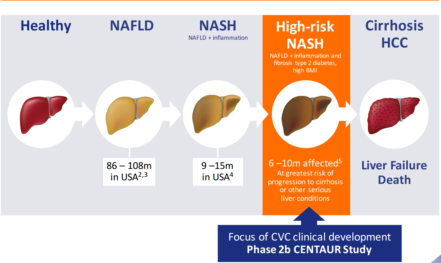
“When MSG is given, mice develop a marked fat deposition within their livers (a type called centrilobular fatty change) leading to fibrosis and hepatic tumors”, a Japanese team of scientists revealed. The changes they observed, showed a marked similarity in microscopic features to non alcoholic fatty liver disease (NAFLD) and non alcoholic steatohepatitis (NASH) in humans.
In addition to above mentioned changes, the mice which received 2 mg/g dose of MSG for 5 days, responded with severe obesity, diabetes mellitus and blood cholesterol abnormalities.
NAFLD and NASH are only two stages of a spectrum of liver diseases, carrying high risk to progress into cirrhosis as illustrated below.

Related image


“When we increased MSG dose given to animals by two fold (4 mg/g), growth rate of all mice suppressed and 10% of the group died. They just couldn’t tolerate it.”, Dr. Nagata, the chief scientist of current study, explained.

Sources:
1.      Nagata M, Suzuki W, Iizuka S, Tabuchi M, Maruyama H, Takeda S, Aburada M, Miyamoto K. Type 2 diabetes mellitus in obese mouse model induced by monosodium glutamate. Exper Animals 2006; 55 (2) 109–115.

2.      A Studies On Monosodium L- Glutamate Toxicity In Animal Models- A Review. Kumar Ganesan1, Kumeshini Sukalingam1, Kanaga Balamurali1, Siti Radziah Bt. Sheikh, Alaudeen1, Kumar Ponnusamy1, Indang Ariati Ariffin, Sharmila Banu Gani. International Journal Of Pharmaceutical, Chemical And Biological Sciences, 2013, 3(4), 1257-1268.


UMAMI - THE NEVER DYING TASTE


MSG is becoming increasingly popular. At the beginning of the century, production of MSG totalled around 1 million tons per year, but it is currently around 3 million. China has taken over from Japan as the major producer, accounting for around 70% of total production. China is also the biggest consumer, but exports have been rapidly increasing in recent years. In 2018, Chinese exports of MSG surpassed 500,000 tons for the first time. Around 90% of the world’s consumption is in Asia, where the increase in MSG demand can be attributed to the higher consumption of ready-meals, as well as increasing disposable incomes.


So we can say that, at least in Asia, the appetite for umami is undiminished. In the West, where glutamates hide in plain sight, we could argue that westerners are equally addicted to that delicious flavour. After all, umami is at stake, but at cost of an array of toxic effects.
(source: http://spendmatters.com/2014/02/03/msg-worlds-well-known-secret-ingredient/)

Image result for asia MSG use

MSG - THE FOOD WORLD'S SECRET

MSG is the most commonly used flavor enhancer after salt and pepper. Glutamic acid  naturally occur in many foods like parmesean cheese, meat, mushrooms and many other foods.

MSG was a discovery of Japanese professor - Kikunae Ikeda in 1908. He extracted this strong flavor enhancer from 'Kombu' sea weeds. As it could be kept as a solid at room temperature, this "umami" (means delicious) taste source became increasingly popular,

Nowadays, it is difficult to find a food manufacturer who avoid use of MSG. Food giants love this, as it greatly enhance flavor and customer satisfaction of any given food making sure a permanent consumer base. Fast food chains are using this special substance for this purpose.

However, with rising public concerns regarding it's unhealthy effects, food giants tend to hide MSG behind various labels such as hydrolyzed vegetable protein, glutamic acid or yeast extracts.

Related image

(photo: Kombu seaweed)

Find more details on MSG in food labels from below links:

http://hapicureclinic.blogspot.com/2018/08/msg-labels-8-natural.html
http://hapicureclinic.blogspot.com/2018/08/msg-labels-7-organic.html
http://hapicureclinic.blogspot.com/2018/08/msg-labels-6-no-msg.html
http://hapicureclinic.blogspot.com/2018/08/msg-labels-5-hydrolyzed-proteins.html
http://hapicureclinic.blogspot.com/2018/08/msg-in-labels-4-they-work-together.html
http://hapicureclinic.blogspot.com/2018/08/msg-in-labels-3-aware-if-you-are-msg.html
http://hapicureclinic.blogspot.com/2018/08/msg-in-labeling-2-its-often-there.html
http://hapicureclinic.blogspot.com/2018/08/msg-in-labels-1-its-always-there.html
http://hapicureclinic.blogspot.com/2018/08/msg-in-diary-products.html


source - http://spendmatters.com/2014/02/03/msg-worlds-well-known-secret-ingredient/


DIABETES RISE IN CHINA, LINKED TO MSG


China is the top producer as well as the top consumer of MSG. In 2014, they have consumed nearly 60% of global consumption of MSG.

 Image result for World MSG consumption

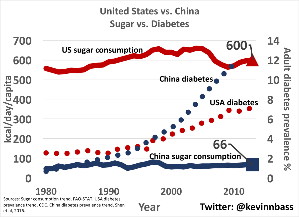
It was clearly mentioned in scientific literature regarding the connection of MSG consumption and increasing diabetic prevalence. Following graph demonstrate the static per capita sugar consumption in China over a 30 year period against skyrocketing diabetes prevalence. Also, note that even U.S. sugar consumption does not show close correlation with diabetes rates.


Related image

Below graph looks into, how MSG production in China closely correlate with China diabetes rise in question. Do you see any connection?

(Two graphs are not drawn in same time frame, but carefully compare 2005 - 2010 steep rise in diabetes in first graph with 2005 -2010 MSG production in second. note that 90% of world MSG production and 60% of world MSG consumption occur in China).

MSG

MSG INDUCE DIABETES IN 5 DAYS


“MSG, if administered at doses of 2 mg/g dose, can produce all the signs of diabetes, along with pathological changes in pancreas, within just 5 days”, researchers say. 

They observed following biochemical changes in mice models of MSG induced diabetes. “These changes were not different from changes occur in diabetes in humans, at all”, they explain.

Biochemical changes include;
  • ·         Severe obesity
  • ·         Urinary glucose
  • ·         Hyperglycemia
  • ·         Hyperinsulinemia
  • ·         Decrease in glucose tolerance
  • ·         Decrease in insulin sensitivity

These experimental animals were developed a severe hypertrophy of pancreatic islets due to excessive beta cell proliferation. (Animal’s and human’s insulin hormone is secreted by beta cells of pancreas). 

So, this method is used by researchers to make animal models diabetic within a short time, while humans continue adding tons of this debatable chemical, into their food chain.

Image result for Diabetic patient

Sources:
1.      Bunyan J, Murrell EA and Shah PP. The induction of obesity in rodents by means of monosodium glutamate. Br J Nutr 1976; 35 (1): 25–39.

2.      A Studies On Monosodium L- Glutamate Toxicity In Animal Models- A Review. Kumar Ganesan1, Kumeshini Sukalingam1, Kanaga Balamurali1, Siti Radziah Bt. Sheikh, Alaudeen1, Kumar Ponnusamy1, Indang Ariati Ariffin, Sharmila Banu Gani. International Journal Of Pharmaceutical, Chemical And Biological Sciences, 2013, 3(4), 1257-1268.


Saturday, August 11, 2018

NATURAL GLUTAMIC ACID vs PROCESSED MSG

Natural glutamic acid


  1. is present in foods such as fish, vegetables, mushrooms, meat, poultry and grains. unprocessed, unadulterated, unfermented proteins in food naturally contain glutamic acid, NOT MSG.
  2. L - glutamic acid is present in nature.
  3. usually present in foods bound to proteins - freed with  digestion of food in intestine.
  4. free glutamic acid naturally present in fermented foods.
  5. need of protein digestion to release amino acids, results in slow absorption - making it easy to be handled by the body.


Synthetic MSG


  1. is produced chemical plants has impurities such as nitroso amines.
  2. Before 1957, MSG was basically extracted from natural protein. some MSG is still produced using this method.
  3. But, majority of MSG today produced (post 1957 era) using genetically modified bacteria.
  4. D- glutamic acid present in synthetic forms.
  5. it's refined - no need of digestion process in body to enter into circulation - making it difficult to be handled by body mechanisms - increases the possibility of MSG toxicity.
  6. MSG sensitive people and vulnerable groups are at high risk


Image result for D glutamic acid in MSG

https://slideplayer.com/slide/8578909/

http://www.truthinlabeling.org/WhatIsMSG.htm


‘MSG IS NOT TOXIC’ – OR IS IT?


‘MSG is not toxic’ – this is a “face book” post that was brought into my attention by a colleague, about 1 week back. “Is that true?”, she asked.

The situation of general public is not different at all. Many are getting confused by number of contradicting evidence that they come across. This is what Glutamate industry needs indeed. They want you to get confused – to keep you in the dark – and away from the truth.

Let’s think of it in a different approach. If you are to call something ‘toxic’, what impression should it generate in your mind?

“I say something toxic, if it could kill me” – that was the answer of my friend. People have a perception in their minds, according to which if something could kill them, they call it toxic. The perception is stronger, if it kills you instantly. If it takes a long time to kill you, you may not aware of it’s true danger.

OK – Let’s go further.
·         Refined sugar, if consumed for long enough in large amounts, would cause diabetes and metabolic syndrome. It is also known as the number one carcinogen in world. Do you call it toxic?
·         Table salt, if consumed in a similar way, would cause hypertension. Do you call it toxic?

Although, we don’t call sugar and salt toxic in everyday life, we do know that they give rise to toxic metabolic effects in long term use. These are “SLOW TOXINS”. They will kill you, but takes a long time for it.

Now, think about MSG. It affects almost every known organ system in human body;
·         It acts as an endocrine disruptor.
·         It can cause metabolic syndrome (a collective term to identify the package of obesity, diabetes, hypertension and dyslipidemia).
·         It gives rise to NAFLD (Non Alcoholic Fatty Liver Disease).
·         It operates as a neurotoxin to dysfunction the ‘master gland’ of hormone control system (hypothalamus), leading to a system breakdown.
·         It can cause retinal degeneration and make you blind.
·         Byproducts of processed MSG can give rise to certain cancers (breast, colon, prostate, pancreatic etc.)
·         It cause ALS, Parkinsonism and Alzheimer’s disease.
·         It causes male and female sub-fertility.
·         It causes thyroid dysfunction and many more.

And it takes a long time to cause any of above. ‘Taking a long time to exert the toxic effect’ cannot be taken as a feature of ‘non toxic’ substance. In summary, it acts as a “SLOW TOXIN”, similar to sugar.

So, what makes it different from sugar?

The answer lies in it’s ability to cause toxic effects in a shorter time frame among vulnerable groups.What are they?
·         Pregnant women
·         Lactating women
·         Women with fertility wishes
·         Pre pubertal children
·         Individuals with liver dysfunction
·         Individuals with micronutrient (specially, Zn, Mg, Vit. B6) deficiencies
(For example, alcoholics, athletes with poor nutrition)

What happen if these people continue to consume MSG?
·         Pregnancy & lactation – 2% of Indian population
a)      Cause maternal overweight/ obese and maternal metabolic derangements.
b)      Reduce weight and length of offspring
c)      Reduced growth rate offspring
·         Women with fertility wishes – 1 in 6 couples are subfertile
a)      Increased miscarriage rate
b)      Reduced implantation of embryos
·         Infants, toddlers and Pre pubertal children – 13%
a)      Destroys memory system
b)      Reduces  learning abilities
c)      Reduces behavioral control
d)     Damages important brain areas to lose appetite control
e)      Destroys interconnections between neural networks reducing intelligence
·         Liver disease patients and people with chronic diseases such as diabetes with various levels of liver dysfunction – liver disease prevalence in India: 2.44% & diabetes prevalence in India: 7.3%
a)      Increases metabolic derangement
b)      Increases disease complications
·         Alcoholics – represent 30% of males and 5% of females in Indian population.
a)      Increases metabolic derangement
b)      Increases disease complications
·         People with mental health disorders – 3% in Indian population

Now sum up the numbers – collective count is around 48%.
If nearly 50% of population is vulnerable to short term damage of MSG, can you call it non toxic?

Certainly NO.

MSG IS TOXIC.
Image result for MSG IS TOXIC